Mating habits, Ovulation, Cleavage
and Development of eggs in Prawns
Copulation takes place in the prawns only
in the night. Prior to copulation the prawn creep about at the bottom,
only one male following a female. In a few minutes the female moults becoming
soft, the male remaining hard without molting. After moulting, the male
advances and embraces the female on the ventral side and they swim with
their bodies inclined. Copulation lasts for 3 or 4 minutes after which
they separate without caring for each other. During copulation the male
ejects the spermatophore containing the sperm through the petasma located
on the fifth pereiopods into the thelycum of the female and the spermatophores
finally reach the seminal vesicle. The same male cannot copulate twice
in the same night because all the spermatophores are deposited in one female.
Spawning takes place at night while swimming
leisurely in the water, one to two feet above the bottom. It begins when
eggs have matured and the germinal vesicle of the egg melts. During spawning
the pereiopods are held tightly to the body and pleopods are moved to and
fro vigorously. Due to this the eggs are scattered behind.
Nearly 700,000 eggs are shed in 3 to 4 minutes
by a prawn of size 20 cm long. The eggs are slightly heavier than
water and if the water is disturbed sinking of eggs is delayed. Spawning
does not weaken the prawn. While the eggs are extruded the spermatozoa
from the spermatophores lodged in the seminal vesicle also extrude out
into the sea water through a minute pair of openings at the base of the
fourth pair of pereopods of the female, and fertilization takes place in
the sea.
The egg inside the ovary is covered with
a layer of follicle cells and possesses 2 or 3 nucleoli. There is no vitelline
membrane. When egg grows it absorbs the follicle cells and in a mature
egg the latter grow smaller becoming a thin membrane, The nucleoli also
becomes smaller and increasing in number. After, all the follicle cells
are absorbed, a jelly-like substance is found on the surface cytoplasm.
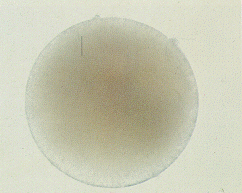
The egg is an irregular sphere before fertilization discharging radially
the whitish, transluscent,jelly-like substance from within. The latter
becomes granular and separates from the egg 7 minutes after spawning, but
a little may remain even after the formation of the fertilization membrane.
By the time the first segmentation is over the jelly may completely disappear
and the egg becomes spherical.
During fertilization a few spermatozoa,
reaches the egg surface one minute after spawning, and after the
jelly is almost disappeared. At the place of contact of the spermatozoa
on the egg, entrance cones appear according to the number of spermatozoa
that have reached the egg. The cytoplasm of a cone protrudes up to the
head of the spermatozoon and into this the head sinks. Soon the cytoplasm
shrinks and drags the spermatozoan into the egg through the egg surface.
Only one spermatozoan is able to pass into the egg even if more sperms
have reached the surface. The others are discarded because the cytoplasms
in their respective entrance cones do not reach up to the heads. When the
sperm is entering the egg the first polar body is extruded before the fertilization
membrance is formed. After this the fertilization membrance is formed around
the egg and the second polar body emerges at the same place as the first
polar body. Finally, the first polar body remains above the membrance,
and the second below it before the first cleavage.
 |
|
An egg soon
after it is spawned
|
The first cleavage is total and equal and
takes place 30 to 40 minutes after spawning. After this further segmentation
takes place every 12 to 15 minutes. After the second division four cells
are formed and when by further cleavage 64 to 128 cells are formed an invagination
occurs at the vegetative cells of the embryo. Soon an embryonic membrane
surrounds the embryo. After about 2 hours and it gradually becomes oval.
The rudiments of the antenna first appears
as a swelling in the middle part of the embryo. The root of the mandible
appears below that of the antenna followed by the swelling of the antennule
above it. When the three pairs of appendages have well developed
the embryo become a nauplius . A dark red ocellus appears on the anterior
end of the body which is ventrally placed .The embryo begins to show signs
of movement within the egg-membrane and in 13 to 14 hours after spawning
the nauplius emerges from it.
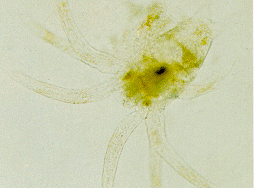
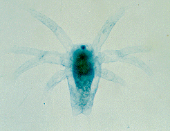
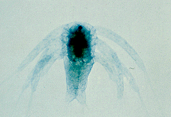
The newly emerged nauplius LARVA has usual
3 pairs of appendages: the anterior uniramous antennules, middle antennae
and posterior mandibles, both biramous. It swims by means of the appendages,
the antennae being the most active, and the antennules coming next. They
are attracted to light but avoid direct sunlight. There are six naupliar
stages, each stage formed after the moulting of the previous stage, their
size ranging from 0.34 mm in the first to 0.51 in the sixth stage. After
each moult body size increases and structural complexities take place.
When at rest, the nauplius keeps its dorsal side down, remaining in water
in a perpendicular position with the three pairs of appendages slanting
upwards. While hatching the colour is dark yellow, later by moulting the
colour becomes whitish and semi-transparent. Appendages get some reddish-
brown specks. Nauplii are not feeding stages, there being yolk in the body
for nourishment. When the protozoea stage is formed the yolk is almost
completely absorbed
 |
|
4th Stage
|
 |
|
5th stage
|
|
|
6th stage
|
The sixth nauplius moults in about 36 to 37
hours and the first protozoea emerges. The body is elongated and changes
are seen in the appearance and swimming behaviour. The endopodites and
exopodites of mandibles fall off, their function now being mastication.
The first and second antennae are the chief organs of locomotion and they
are also aided in swimming by the first and second maxillipeds which have
developed well. It is photopositive but shuns very bright light. The carapace
covers the anterior side of the body up to the eighth segment. The compound
eyes make their appearance towards the end of this stage. The uncovered
eyes make their appearance towards the end of this stage. The uncovered
lower part has six segments which belong to the thoracic region. Later
five abdominal segments develop with indistinct boundary lines. The telson
is well developed with a semi- spherical forked end. The appendages are
well developed with distinct articulations and plumose setae and spines.
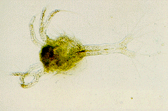
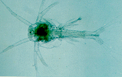
There are three protozoea stages formed
by moulting, and after each moult more appendages and other structures
are formed. Eye protrude out in the second protozoea stage. The third protozoea
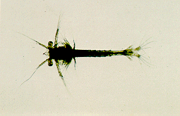
moults into the mysis stage. The mysis resembles the adult prawn
having the cephalic and thoracic segments united to form a cephalothorax
which is covered by the carapace. Rostrum develops to a little more than
half the length of the carapace. The first and second antenna cease to
be natatory and become olfactory in function. The five pairs of pereopods
take on the function of swimming, the three pairs of maxillipeds also assisting.
They swim with their heads down and telson up, keeping the body in a slanting
position. They do not show much preference to light. The body colour is
pale yellow, the colour of the mouth parts, thorax, abdominal segments
and tip of first antenna is reddish brown.
 |
|
A zoea (after
3 molting the Zoea transforms into a Mysis)
|
 |
|
2nd Stage
|
 |
|
3rd Stage
|
The size of the mysis stages varies from about
3.10 mm length in the first to 4.52 mm in the third stage. The abdomen
is a little shorter than the body. On the basal segment of the antennule
with three segments the rudiment of an otolith is developed. The rudiments
of the green gland appear on the protopodite of antenna. On the first three
pairs of pereopods of early mysis, rudiments of arthrobranchs appear and
they become clearly distinguishable in the third mysis. In this stage a
rudiment of arthrobranch appears on the fourth pair of pereopods also.
The five pairs of pleopods on abdomen are rudimentary and functionless.
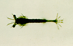
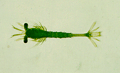
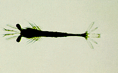
There are three mysis stages formed by moulting. After each molt the body
structure change. The third mysis moults into the first postLARVAe.
 |
|
A Mysis (After
3 moltings, the Mysis transforms into a Post LARVA)
|
 |
|
2nd Stage
|
 |
|
3rd Stage
|
In the postLARVA the five pairs of pleopods
fully developed and functions as the chief organ for swimming, the uropods
also assisting in balancing. The pereopods are used only for walking and
grasping. At this stage it goes down and creeps on the sand below, which
was still now swimming in the water column. Day and night they creep and
burrow into the sand at intervals. After 10 or 12 moults they begin to
creep on the sand as the adults. After 20 to 22 moults the shape of the
body and appendages resemble those of the adults. The colour changes in
the postLARVA after four or five moults to greenish-black, brownish-black,
brownish-yellow etc on various part of the body.
POST LARVAL
STAGES.
Copyright 1998 Bioinformatics Centre,
National Institute of Oceanography, Dona Paula, Goa, India